ODM Thoracolumbar Interbody Cage (Angled) Exporter, Company
Product Description
| Material | PEEK |
|---|---|
| Qualification | CE / ISO13485 / NMPA |
| Package | Sterile Packaging, 1 pcs/package |
| MOQ | 1 Pcs |
| Supply Ability | 1000+ Pieces per Month |
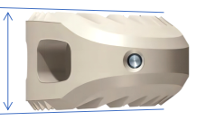
| Available Dimensions (28 mm Length) | 7 mm, 8 mm, 9 mm, 10 mm, 11 mm, 12 mm, 13 mm, 14 mm Height x 28 mm Length |
| Available Dimensions (31 mm Length) | 7 mm, 8 mm, 9 mm, 10 mm, 11 mm, 12 mm, 13 mm, 14 mm Height x 31 mm Length |
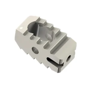
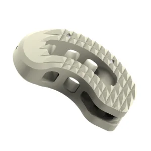
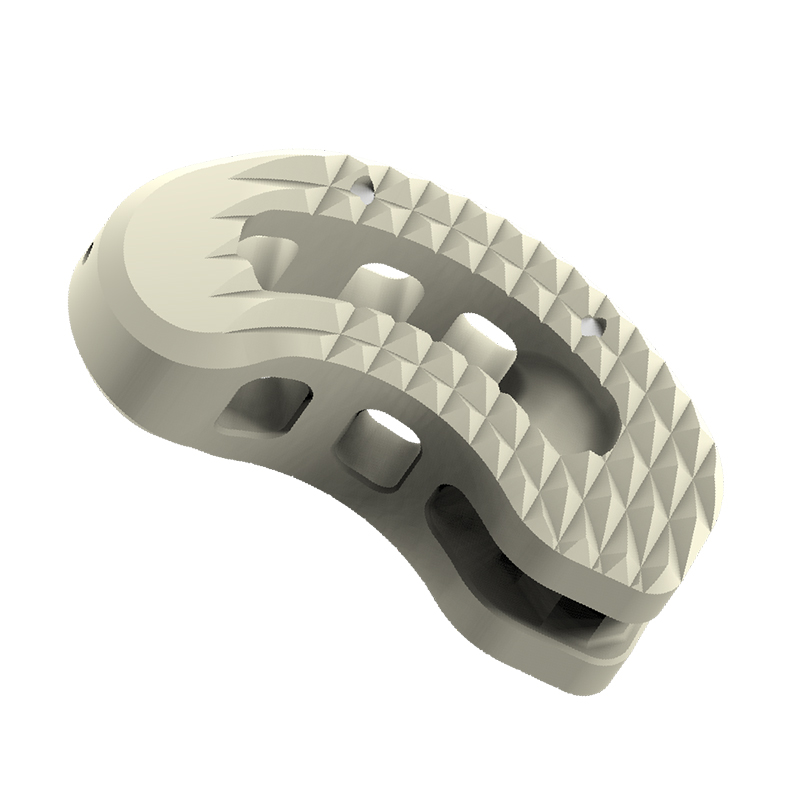
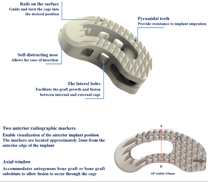
Provide resistance to implant migration.
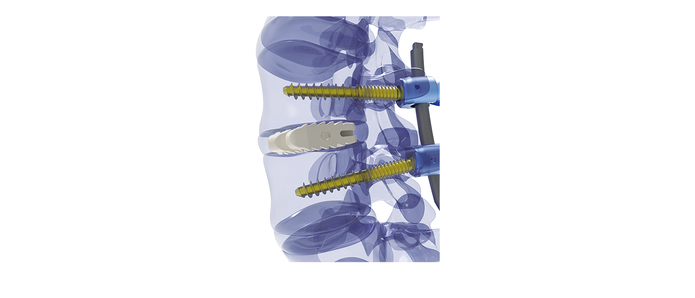
Allows for ease of insertion during entry.
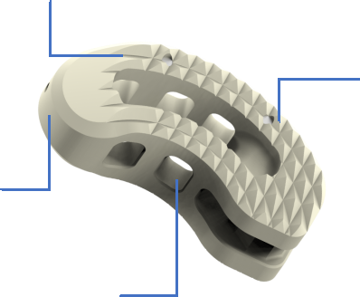
Guide and turn the cage into the desired position.
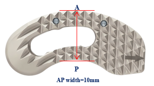
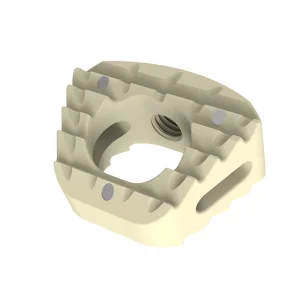
Two anterior radiographic markers: Enable visualization of the anterior implant position (located approximately 2mm from the anterior edge of the implant).
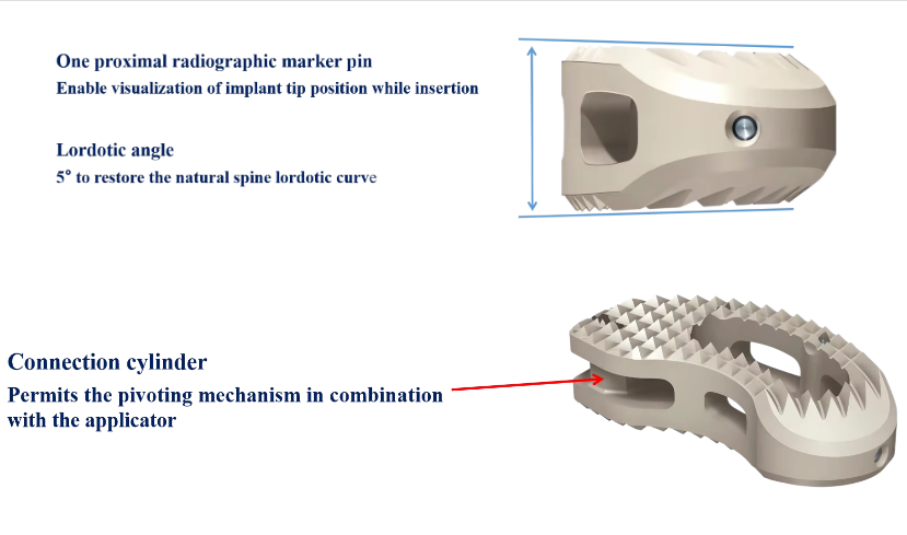
One proximal radiographic marker pin: Enable visualization of implant tip position while insertion.
Lordotic angle 5°: Engineered to restore the natural spine lordotic curve.
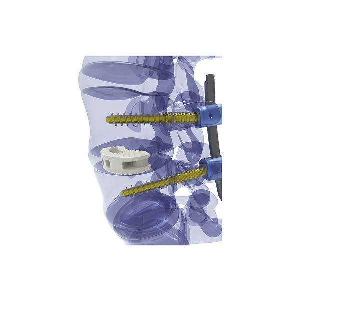
Axial window: Accommodates autogenous bone graft or bone graft substitute to allow fusion to occur through the cage.

The lateral holes: Facilitate the graft growth and fusion between internal and external cage.
Connection cylinder: Permits the pivoting mechanism in combination with the applicator.
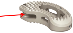
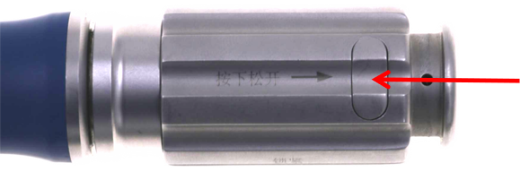
• One key instrument for insertion of implant and trials.
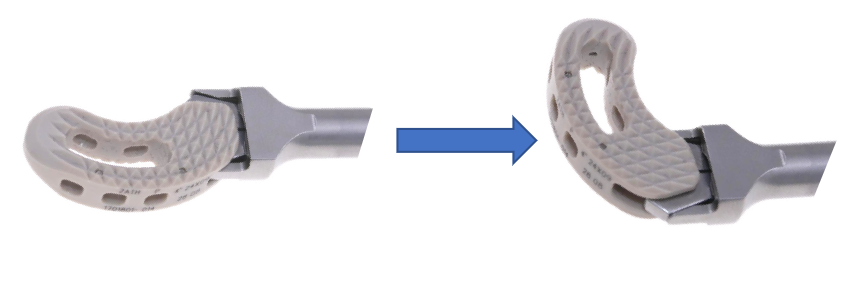
• Applicator allows a controlled and guided insertion based on the pivoting option.
• Security button to prevent implant disengagement.
• Applicator is designed for minimally invasive surgeries.
• Disassemble button for easy cleaning.
Engineered for precise angle navigation and secure coupling during minimally invasive spine procedures.
The cage is manufactured from high-grade medical PEEK (Polyetheretherketone), which offers an elasticity modulus close to human bone and provides radiolucency for unimpeded radiographic evaluation.
The cage comes in lengths of 28 mm and 31 mm, with heights ranging in 1 mm increments from 7 mm to 14 mm. It features a 5° lordotic angle to restore the physiological curvature of the spine.
It includes two anterior radiographic markers situated approximately 2 mm from the anterior border and one proximal radiographic marker pin at the tip, enabling clear fluoroscopic visualization throughout insertion.
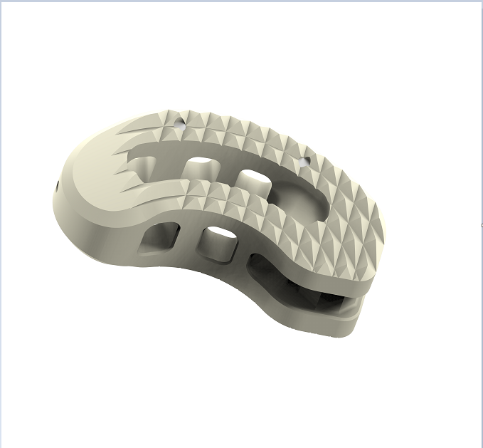
Pyramidal teeth grip adjacent endplates to resist migration. A spacious axial window accepts autogenous bone graft or bone graft substitute, while lateral holes facilitate osseous integration and fusion between the inside and outside of the cage.
The dedicated applicator functions with both trials and implants, utilizing a connection cylinder for a controlled pivoting insertion. It incorporates a security button to avoid accidental disengagement and a disassemble button for simplified hospital cleaning and maintenance.
The implant holds CE, ISO13485, and NMPA certifications. Each implant is supplied individually in sterile packaging (1 pcs/package) with an MOQ of 1 piece and a supply capacity exceeding 1,000 pieces per month.